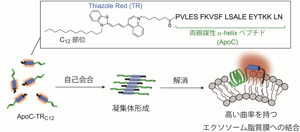
Exosomes, a subgroup of extracellular vesicles that generally range from 30 to 150 nm in diameter, hold great potential in diagnostic application. In order to understand their biological functions, it is of great importance to develop analytical methods for rapid, simple and sensitive detection of exosomes. Here, we developed a new class of fluorescent probes targeting exosomal membranes to this aim [1]. Amphipathic helical (AH) peptides capable of selective binding to highly curved membranes (C-terminal region of apolipoprotein A-1: ApoC)[2] were coupled with lipophilic tail-carrying cyanine dyes (TRC12)for the fluorescence detection of exosomes based on the binding-induced fluorescent response. ApoC-TRC12 has extremely weak emission due to the formation of the aggregates, whereas it becomes emissive in response to the target exosomes through the binding-induced disassembly of ApoC-TRC12. We demonstrated the useful function of ApoC-TRC12 for rapid, simple and sensitive detection of exosomes.