The primary cilium is a microtubule-based tiny organelle emanating from the surface of most eukaryotic cells. Cilium receives various chemical and mechanical signals from the extracellular milieu and plays critical roles in the development and homeostasis of many organ systems. Defects in cilium formation or function cause diverse genetic disorders, called ciliopathies. It is difficult to analyze the structure of primary cilia because of the size approximates the optical diffraction limit (approximately 0.3 µm in diameter and 3 µm in length).
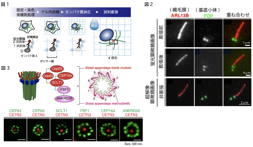
Expansion microscopy (ExM) is an alternative super-resolution imaging technique that uses a swellable hydrogel that enables the physical expansion of specimens. In this study, we applied ExM to observe primary cilia and centrioles and compared the acquired images with those obtained using conventional super-resolution microscopy. We also demonstrate that the combinatorial use of ExM protocol with super-resolution microscopy, enables the practical observation of primary cilia and basal bodies with high resolution.